Honors Thesis
Assessing Endothelial Cell Compatibility in Hydrogels for 3D Coaxial Bioprinting of Patterned Vasculature
My Impact & Learned Skills
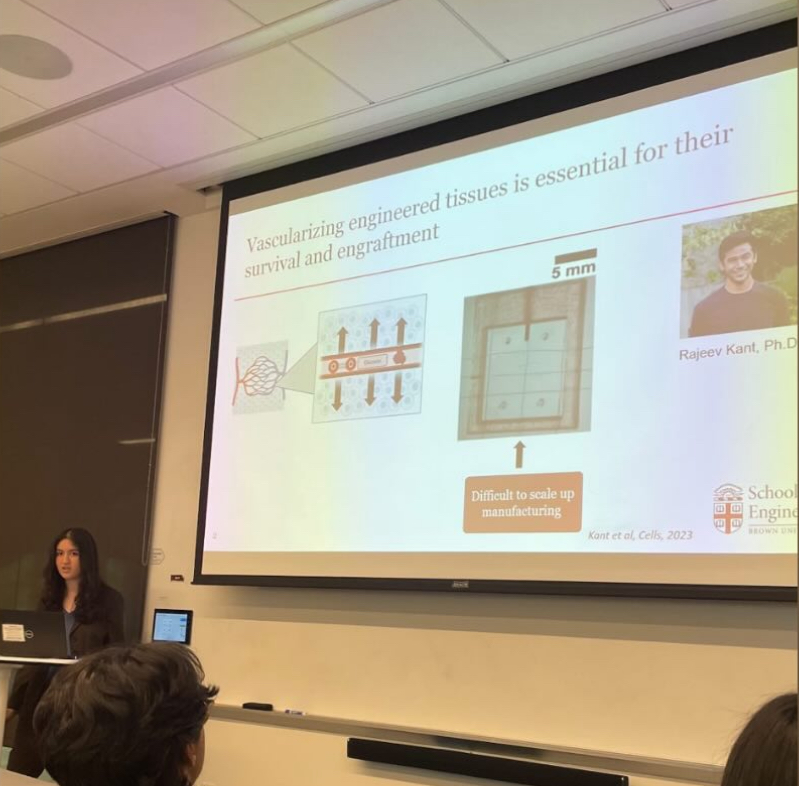
Presenting my Work at the Brown Engineering Honors Thesis Symposium
Throughout this project, I grew my foundational skills in biomedical research and learned to become an independently driven researcher. I practiced methods such as cell culture, microscopy, fluorescent staining, and aseptic technique. My thesis was an important development in my lab’s knowledge surrounding vascular engineering and has yielded promising data about using this hydrogel for future research. My work moves the field of vascular and tissue engineering forward and has taught me about the current state of the field. I’ve practiced experimental design, especially through the creation of assays for each specific characteristic I wanted to measure in my studies. I’ve learned how to conduct interdisciplinary research as my research combined cell biology knowledge with key engineering concepts. I was able to combine knowledge from coursework across multiple areas of study to understand how to quantify experimental data and set up experimental conditions. I’ve learned to put statistical methods into practice to analyze and visualize key findings from my data. I’ve also written scientific manuscripts and communicated my thesis to a broader audience effectively via public presentations. My time in the Coulombe Lab has been essential to my development as a researcher and has grown my motivation for pursuing science as a career to create future health innovations.

Bioprinting Workflow to Engineer Vasculature in Synthetic Tissue
I spent two years in the Coulombe Lab for Heart Health and Regeneration working on an independent research project to bioprint vasculature (blood vessels) for implantation in synthetic tissues. Using a technique called core-shell or coaxial bioprinting, two concentric cylinders can be printed simultaneously, containing two different materials. The inner layer, known as the core, would be a sacrificial material that could be hardened through a process called cross-linking and then chelated or un-cross-linked to create a hollow channel surrounded by the shell. Using various biocompatible polymers called hydrogels, I optimized a unique formulation for the shell layer where the endothelial cells (ECs, cells that line all blood vessels) would be located. Using biomaterial engineering practices, I assessed this formulation for EC compatibility and structural properties. The ideal material must support cells through the printing process, amidst extrusion, cross-linking, and chelation. My thesis focused on studying cell performance through properties like proliferation, viability, and adhesion amidst these processes to assess the cell compatibility of the gel, called GAF.
.png")
This project addresses a key problem in current tissue engineering. As engineered tissues grow in complexity to solve greater problems in regenerative medicine – such as whole organ replacement – they must be pre-vascularized with engineered vasculature. In native tissues, vasculature provides essential ingredients for life such as oxygen, glucose, proteins, and other key nutrients. Without a constant supply of these ingredients, tissues can have poor survival outcomes. Therefore, having a vasculature supply is key to the long-term survival of any tissue, and engineered tissues must contain vasculature to approach biomimicry, survive effectively, and successfully engraft with their hosts.

Fluorescent Imaging Used for Live-Dead Cytoticity Assays
.jpg")
Proliferation Assay Using Cell Staining Dye over Multiple Days
Throughout my thesis, I designed assays to measure these key characteristics of cell compatibility over various timelines to mimic the bioprinting workflow. This included making a simulatory bioprinter using a syringe pump and needles, performing both 2D and 3D experiments, and optimizing experimental conditions with each iteration to better approach the true workflow conditions. I performed fluorescent imaging for most of my assays, using this visual data to create quantitative metrics of cell viability, proliferation, and adhesion for each group. My time in the lab, especially while doing my thesis work, taught me how to perform independent research, gain quantitative metrics of experimental data, design effective experiments, and persevere through challenges when unexpected results were obtained. I’ve learned a lot about how engineering concepts can be combined with biomedical research to create new technology. My thesis ultimately showed that our GAF gel was a promising candidate for this application, opening the doors for actual vascular fabrication in the future.